
Motion movies have evolved phenomenally for decades, giving us more real videos than waved and blurred videos that we all watch. Evolution in practice is caused by advances in imaging technology for many years, starting with a simple video ~ 16 frames per second (FPS) by Lumiere Brothers 18th century into a deep game environment with ~ 150 fps. In addition to entertainment, can we use this technology to imagine and understand the microscopic work of the human body complex, such as neuron activity in the human brain?
In innovative steps taken by photonic and quantum that allows the sensing of the Technology Laboratory, Indian Bombay Institute of Technology, Laboratory Leader Prof. Kasturi Saha and his team exploit the capabilities of high FPS cameras specifically to get images that change time or ‘dynamic’ from a very weak magnetic field, Typical of what is found in neurons. A study based on this work published in the journal Scientific Reports.
We use neuroimaging techniques such as Magnetic Resonance Imaging (MRI), Magnetoencephalography (Meg) and Functional Magnetic Resonance Imaging (FMRI) to study the internal structure of the human brain. These techniques measure electromagnetic signals that provide snapshots of combined neuron activity and help us study certain brain functions. In addition, these signals are weak, so we need a large and very strong magnet in this technology to detect it. Thus, how to increase our ability to detect weak neuron signals using efficient technology is one of the urgent questions answered by the Neurologist and Medical Community in general.
Often, we find imperfections in the structure of the diamond crystal lattice, one of which includes nitrogen atoms located next to the vacancies in the diamond grid. The center of nitrogen-vacancy (NV) defects has ‘non-paired’ electrons that are very sensitive to external triggers such as magnetic fields and temperatures, making it a unique quantum probe for measuring weak but very sensitive magnetic fields at the microscopic level. This change can be measured directly by feeling low intensity light, also known as ‘photoluminescence’, which is emitted from the interaction of these electrons with a magnetic field. Researchers take advantage of the unique properties of this NV to a very sensitive magnetometer, the size of the atom that can map the magnetic field on a smaller physical scale.
The neuronal magnetic field changes faster than how quickly the current NV -based magnetometer can capture changes. With the current NV magnetometer, arrest takes a few minutes to a few hours for high resolution images, and therefore whatever changes faster than that cannot be visited and basically only gives us snapshot. Prof. Saha and his team are the first research groups to present a unique and efficient unique increase using a ‘lock-in’ camera that is available specifically in the magnetic field microscope setting that reduces the acquisition time from a few minutes per frame to the order of 100 fps. Globally this is the first time anyone has shown a sub-second magnetic field microscope with this technique.
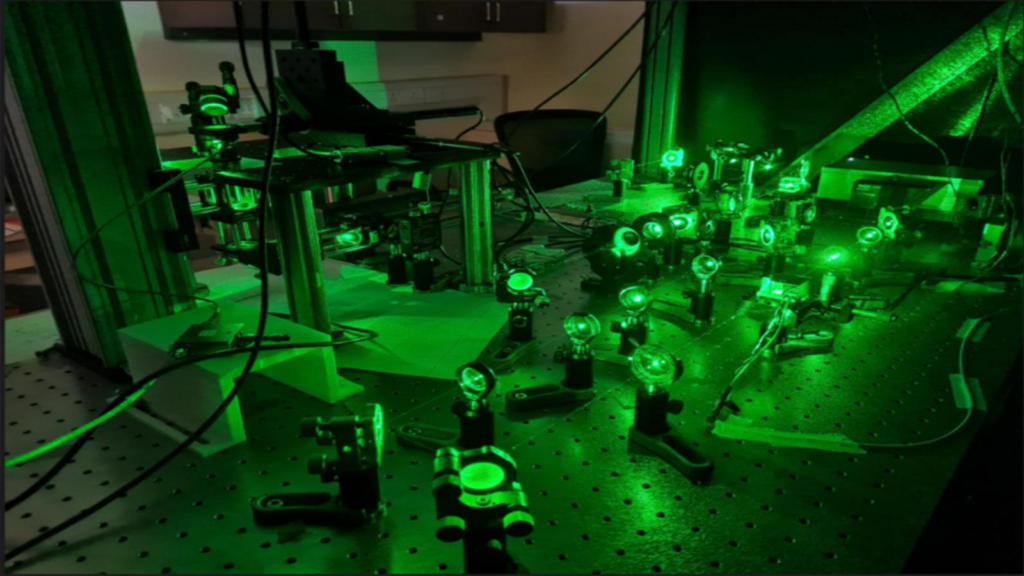
They set the experiment by installing pure diamond crystal sheets that were very thin with NV defects in two types of magnetic field sources – very thin microscopic wire (or ‘microwire’) and microscopic wire coils (or ‘microcoil’). They introduce variations in a magnetic field by manipulating the central rotation of the NV with a micro wave frequency that changes on the millisecond scale. This rapidly changing magnetic field is captured through the interaction at the NV defect center in a diamond crystal layer, which produces low light photoluminescence.
However, the ingenuity of this experiment lies in how the Prof. Saha team uses a high FPS locking camera to capture the changing magnetic field. Unlike the conventional camera, in a locked camera, each pixel only detects light fluctuations that contain specific frequencies or oscillations from a fixed period of time and ‘reject’ light fluctuations from other frequencies. This increases the signal for the locked camera and therefore, suitable in very low light imaging scenarios such as photoluminescence from the center of NV. As a result, they can get a ‘movement image’ from a magnetic field from Microwire and Microcoil by capturing any changes in the magnetic field by adjusting the locking camera to capture the lamp emitted by the NV center.
NV -based microscope thus has the potential to become a promising alternative technology to investigate mammalian brain activity. How to encourage the accuracy of the sensing of electromagnetic fields at the microscopic level using the quantum properties of atomic constituents forming the core of the new fields that will come from ‘quantum sensing’.


More Stories
NASA posts beautiful martian image showing ‘blue’ region of the red planet
NASA posts beautiful martian image showing ‘blue’ region of the red planet
Formation of dwarf galaxy observed using India’s AstroSat